Ever wondered how scientists can see tiny cells and discover...
Microscope Review for Beginners: Understanding Magnification
gift@esilia
1
of 2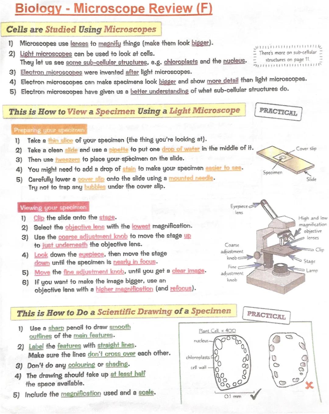
Using Microscopes to Study Cells
Microscopes are basically powerful magnifying glasses that let you see things way too small for your naked eye. Light microscopes are brilliant for studying cells and can show you cool structures like chloroplasts and the nucleus inside cells.
Electron microscopes are the supercharged versions that came later. They can magnify specimens much more than light microscopes and reveal incredible detail. These high-tech tools have revolutionised our understanding of what actually happens inside cells.
When preparing a specimen for viewing, you'll slice it thin, place it on a slide with a drop of water, and might add some stain to make features pop out. The trick is lowering that cover slip carefully with tweezers to avoid air bubbles - they're annoying and ruin your view!
To actually view your specimen, start with the lowest magnification objective lens and use the coarse adjustment knob first, then fine-tune with the fine adjustment. Always look through the eyepiece whilst moving the stage down until everything comes into focus.
Top Tip: When drawing what you see, use a sharp pencil for smooth lines, label clearly without crossing lines, and never colour or shade your scientific drawings!
2
of 2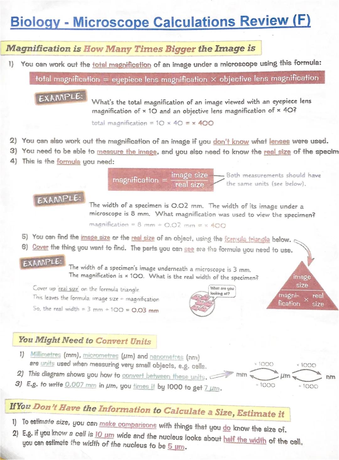
Magnification Calculations Made Simple
Working out total magnification is straightforward once you know the formula: eyepiece lens magnification × objective lens magnification. If your eyepiece is ×10 and objective lens is ×40, you get ×400 total magnification.
Sometimes you'll need to calculate magnification when you don't know what lenses were used. The key formula is: magnification = image size ÷ real size. Just make sure both measurements use the same units!
The formula triangle is your best friend for these calculations. Cover what you want to find, and what's left showing gives you the formula. Need real size? Cover it up and you'll see image size ÷ magnification.
Unit conversions are crucial in microscopy work. Remember that millimetres (mm) convert to micrometres (µm) by multiplying by 1000, and µm converts to nanometres (nm) the same way. Going backwards, you divide by 1000 each time.
Quick Trick: If you can't measure something precisely, estimate by comparing it to something you do know the size of - like saying a nucleus looks half the width of a 10 µm cell, so it's roughly 5 µm!
We thought you’d never ask...
Our AI Companion is a student-focused AI tool that offers more than just answers. Built on millions of Knowunity resources, it provides relevant information, personalised study plans, quizzes, and content directly in the chat, adapting to your individual learning journey.
You can download the app from Google Play Store and Apple App Store.
That's right! Enjoy free access to study content, connect with fellow students, and get instant help – all at your fingertips.
Similar content
Most popular content: Microscope
9GCSE Biology Practical Insights
Explore essential GCSE Biology practicals covering microscopy, photosynthesis, enzyme activity, food tests, and osmosis. This comprehensive guide includes key concepts, experimental procedures, and potential exam questions to enhance your understanding and preparation for Paper 1.
93,496155
Biology Required Practicals Overview
Explore the essential first four required practicals for GCSE Biology, including microscopy techniques, microbiology methods, osmosis experiments, and food testing protocols. This summary provides clear steps and safety guidelines to enhance your understanding and preparation for practical assessments.
102,48432
Microscopy Essentials
Explore the fundamentals of microscopy, including the differences between light and electron microscopes, magnification calculations, and practical usage tips. This summary covers key concepts such as microscope types, magnification formulas, and step-by-step instructions for using a light microscope, making it an essential resource for GCSE biology students.
96516
Cell Structure whole topic + Some biological molecules
OCR Biology A level
122027
Microscope Types & Functions
Explore the various types of microscopes including Optical, Transmission Electron Microscopes (TEMs), and Scanning Electron Microscopes (SEMs). This summary covers key concepts such as magnification, resolution, and the advantages and disadvantages of each microscope type. Ideal for A-level biology students studying cell structure and microscopy techniques.
121,47238
Understanding Light Microscopes
Explore the fundamentals of light microscopes, including their components, magnification calculations, and the size range of animal and plant cells. This summary provides essential insights for biology students studying microscopy techniques and cellular structures.
10680
Microscope Practical Guide
Explore the essential techniques for observing onion and human cheek cells using a light microscope. This practical guide covers slide preparation, magnification calculations, safety measures, and a comparison between light and electron microscopes. Ideal for biology students seeking to enhance their microscopy skills.
113223
Microscope Types & Functions
Explore the fundamentals of microscopy, including the differences between light and electron microscopes, their magnification capabilities, and resolving power. This summary covers key concepts such as image size, magnification calculations, and the significance of resolution in microscopy. Ideal for AQA Biology students preparing for exams.
1036612
Types of Microscopes
Explore the differences between light and electron microscopes, including their structures, magnification capabilities, and applications in viewing cellular details. This summary provides essential insights into how these instruments enhance our understanding of microscopic life.
1144512
Most popular content in Biology
9C
Cell Biology and Cell structure
cell structures
93,2810
T
The functions of subcellular structures - B1 Biology
Flashcards on the different functions of subcellular structures: cell membrane, nucleus, mitochondria, ribosomes, cytoplasm, permant vacuole, chloroplasts and cell wall.
102,8215
1
1.cells Gcse biology question cards
combined science higher biology
92,3644
AQA Biology: Key Concepts
Explore essential AQA Biology topics including Photosynthesis, Respiration, Homeostasis, Genetics, and Ecology. This comprehensive knowledge organizer covers key concepts such as energy transfer, hormonal control, and genetic variation, providing a solid foundation for your studies. Ideal for exam preparation and understanding biological processes.
108,878308
B
Biology Paper 1 quiz
this is a simple quiz on key knowledge needed for paper 1
102,7835
T
Types of cells
biology
101,2170
A-Level Biology Year 1 Overview
Comprehensive summary of AQA A-Level Biology Year 1, covering key topics such as cellular structure, protein synthesis, immune response, gas exchange, and more. Ideal for exam preparation and understanding biological concepts. Includes detailed insights into cellular processes, biological classification, and the circulatory system.
1214,928697
biology paper 1
all notes
102,47937
B
biology paper 1
these are some exam questions for biology paper which are more likely to come up in exams
101,8050
Most popular content
9Sociology of Education Overview
Explore comprehensive A-Level Sociology notes on the education system, covering key theories, policies, and sociological perspectives. This resource includes insights on marketisation, gender roles, cultural deprivation, and educational inequalities, providing a thorough understanding of how education shapes social stratification and individual achievement. Ideal for exam preparation and in-depth study.
12102,9583,041
Sociology of Families: Comprehensive Revision
Dive into an extensive overview of family dynamics, perspectives, and patterns in sociology. This resource covers key concepts such as family diversity, gender roles, marriage, and the impact of social policies on family structures. Perfect for A-Level Sociology students preparing for Paper 2.
1273,7532,307
Criminology: Crime & Punishment Overview
Comprehensive mindmaps covering key concepts in the Crime and Punishment topic for WJEC Criminology Unit 4. This resource includes detailed insights into the Criminal Justice System, crime prevention strategies, sentencing models, and the roles of various agencies. Ideal for A-Level revision, ensuring you grasp essential theories and legislative processes to excel in your exams.
1254,8941,060
Comprehensive Crime & Deviance Overview
Explore an extensive revision of crime and deviance topics, including theories, types of crime, and the impact of media. This resource covers key concepts such as Marxism, functionalism, gender and crime, and the influence of globalization on criminal behavior. Ideal for students seeking a thorough understanding of criminology and its various theories. Type: Full Topic Revision.
1251,6691,400
C
Cell Biology and Cell structure
cell structures
93,2810
WJEC Unit 4 Criminology
Criminology unit 4 detailed revision note
127,176125
Sociological Theories Overview
Comprehensive revision of key sociological theories including Functionalism, Marxism, Feminism, and Interpretivism. Explore concepts like value freedom, identity formation, and the critique of social control. Ideal for AQA A-Level Sociology students preparing for exams. This summary covers essential theories and their implications in sociology, providing a clear understanding of each perspective.
1231,480846
T
The functions of subcellular structures - B1 Biology
Flashcards on the different functions of subcellular structures: cell membrane, nucleus, mitochondria, ribosomes, cytoplasm, permant vacuole, chloroplasts and cell wall.
102,8215
1
1.cells Gcse biology question cards
combined science higher biology
92,3644
Students love us — and so will you.
4.6/5App Store
4.7/5Google Play
The app is very easy to use and well designed. I have found everything I was looking for so far and have been able to learn a lot from the presentations! I will definitely use the app for a class assignment! And of course it also helps a lot as an inspiration.
Stefan SiOS user
This app is really great. There are so many study notes and help [...]. My problem subject is French, for example, and the app has so many options for help. Thanks to this app, I have improved my French. I would recommend it to anyone.
Samantha KlichAndroid user
Wow, I am really amazed. I just tried the app because I've seen it advertised many times and was absolutely stunned. This app is THE HELP you want for school and above all, it offers so many things, such as workouts and fact sheets, which have been VERY helpful to me personally.
AnnaiOS user
Microscope Review for Beginners: Understanding Magnification
gift@esilia
Ever wondered how scientists can see tiny cells and discover what's inside them? Microscopes are your gateway to exploring the invisible world around you. Understanding how to use microscopes and calculate magnifications is essential for biology studies and will help...
1
of 2
Sign up to see the content. It's free!
- Access to all documents
- Improve your grades
- Join milions of students
By signing up you accept Terms of Service and Privacy Policy
Using Microscopes to Study Cells
Microscopes are basically powerful magnifying glasses that let you see things way too small for your naked eye. Light microscopes are brilliant for studying cells and can show you cool structures like chloroplasts and the nucleus inside cells.
Electron microscopes are the supercharged versions that came later. They can magnify specimens much more than light microscopes and reveal incredible detail. These high-tech tools have revolutionised our understanding of what actually happens inside cells.
When preparing a specimen for viewing, you'll slice it thin, place it on a slide with a drop of water, and might add some stain to make features pop out. The trick is lowering that cover slip carefully with tweezers to avoid air bubbles - they're annoying and ruin your view!
To actually view your specimen, start with the lowest magnification objective lens and use the coarse adjustment knob first, then fine-tune with the fine adjustment. Always look through the eyepiece whilst moving the stage down until everything comes into focus.
Top Tip: When drawing what you see, use a sharp pencil for smooth lines, label clearly without crossing lines, and never colour or shade your scientific drawings!
2
of 2Sign up to see the content. It's free!
- Access to all documents
- Improve your grades
- Join milions of students
By signing up you accept Terms of Service and Privacy Policy
Magnification Calculations Made Simple
Working out total magnification is straightforward once you know the formula: eyepiece lens magnification × objective lens magnification. If your eyepiece is ×10 and objective lens is ×40, you get ×400 total magnification.
Sometimes you'll need to calculate magnification when you don't know what lenses were used. The key formula is: magnification = image size ÷ real size. Just make sure both measurements use the same units!
The formula triangle is your best friend for these calculations. Cover what you want to find, and what's left showing gives you the formula. Need real size? Cover it up and you'll see image size ÷ magnification.
Unit conversions are crucial in microscopy work. Remember that millimetres (mm) convert to micrometres (µm) by multiplying by 1000, and µm converts to nanometres (nm) the same way. Going backwards, you divide by 1000 each time.
Quick Trick: If you can't measure something precisely, estimate by comparing it to something you do know the size of - like saying a nucleus looks half the width of a 10 µm cell, so it's roughly 5 µm!
We thought you’d never ask...
Our AI Companion is a student-focused AI tool that offers more than just answers. Built on millions of Knowunity resources, it provides relevant information, personalised study plans, quizzes, and content directly in the chat, adapting to your individual learning journey.
You can download the app from Google Play Store and Apple App Store.
That's right! Enjoy free access to study content, connect with fellow students, and get instant help – all at your fingertips.
Similar content
Most popular content: Microscope
9GCSE Biology Practical Insights
Explore essential GCSE Biology practicals covering microscopy, photosynthesis, enzyme activity, food tests, and osmosis. This comprehensive guide includes key concepts, experimental procedures, and potential exam questions to enhance your understanding and preparation for Paper 1.
93,496155
Biology Required Practicals Overview
Explore the essential first four required practicals for GCSE Biology, including microscopy techniques, microbiology methods, osmosis experiments, and food testing protocols. This summary provides clear steps and safety guidelines to enhance your understanding and preparation for practical assessments.
102,48432
Microscopy Essentials
Explore the fundamentals of microscopy, including the differences between light and electron microscopes, magnification calculations, and practical usage tips. This summary covers key concepts such as microscope types, magnification formulas, and step-by-step instructions for using a light microscope, making it an essential resource for GCSE biology students.
96516
Cell Structure whole topic + Some biological molecules
OCR Biology A level
122027
Microscope Types & Functions
Explore the various types of microscopes including Optical, Transmission Electron Microscopes (TEMs), and Scanning Electron Microscopes (SEMs). This summary covers key concepts such as magnification, resolution, and the advantages and disadvantages of each microscope type. Ideal for A-level biology students studying cell structure and microscopy techniques.
121,47238
Understanding Light Microscopes
Explore the fundamentals of light microscopes, including their components, magnification calculations, and the size range of animal and plant cells. This summary provides essential insights for biology students studying microscopy techniques and cellular structures.
10680
Microscope Practical Guide
Explore the essential techniques for observing onion and human cheek cells using a light microscope. This practical guide covers slide preparation, magnification calculations, safety measures, and a comparison between light and electron microscopes. Ideal for biology students seeking to enhance their microscopy skills.
113223
Microscope Types & Functions
Explore the fundamentals of microscopy, including the differences between light and electron microscopes, their magnification capabilities, and resolving power. This summary covers key concepts such as image size, magnification calculations, and the significance of resolution in microscopy. Ideal for AQA Biology students preparing for exams.
1036612
Types of Microscopes
Explore the differences between light and electron microscopes, including their structures, magnification capabilities, and applications in viewing cellular details. This summary provides essential insights into how these instruments enhance our understanding of microscopic life.
1144512
Most popular content in Biology
9C
Cell Biology and Cell structure
cell structures
93,2810
T
The functions of subcellular structures - B1 Biology
Flashcards on the different functions of subcellular structures: cell membrane, nucleus, mitochondria, ribosomes, cytoplasm, permant vacuole, chloroplasts and cell wall.
102,8215
1
1.cells Gcse biology question cards
combined science higher biology
92,3644
AQA Biology: Key Concepts
Explore essential AQA Biology topics including Photosynthesis, Respiration, Homeostasis, Genetics, and Ecology. This comprehensive knowledge organizer covers key concepts such as energy transfer, hormonal control, and genetic variation, providing a solid foundation for your studies. Ideal for exam preparation and understanding biological processes.
108,878308
B
Biology Paper 1 quiz
this is a simple quiz on key knowledge needed for paper 1
102,7835
T
Types of cells
biology
101,2170
A-Level Biology Year 1 Overview
Comprehensive summary of AQA A-Level Biology Year 1, covering key topics such as cellular structure, protein synthesis, immune response, gas exchange, and more. Ideal for exam preparation and understanding biological concepts. Includes detailed insights into cellular processes, biological classification, and the circulatory system.
1214,928697
biology paper 1
all notes
102,47937
B
biology paper 1
these are some exam questions for biology paper which are more likely to come up in exams
101,8050
Most popular content
9Sociology of Education Overview
Explore comprehensive A-Level Sociology notes on the education system, covering key theories, policies, and sociological perspectives. This resource includes insights on marketisation, gender roles, cultural deprivation, and educational inequalities, providing a thorough understanding of how education shapes social stratification and individual achievement. Ideal for exam preparation and in-depth study.
12102,9583,041
Sociology of Families: Comprehensive Revision
Dive into an extensive overview of family dynamics, perspectives, and patterns in sociology. This resource covers key concepts such as family diversity, gender roles, marriage, and the impact of social policies on family structures. Perfect for A-Level Sociology students preparing for Paper 2.
1273,7532,307
Criminology: Crime & Punishment Overview
Comprehensive mindmaps covering key concepts in the Crime and Punishment topic for WJEC Criminology Unit 4. This resource includes detailed insights into the Criminal Justice System, crime prevention strategies, sentencing models, and the roles of various agencies. Ideal for A-Level revision, ensuring you grasp essential theories and legislative processes to excel in your exams.
1254,8941,060
Comprehensive Crime & Deviance Overview
Explore an extensive revision of crime and deviance topics, including theories, types of crime, and the impact of media. This resource covers key concepts such as Marxism, functionalism, gender and crime, and the influence of globalization on criminal behavior. Ideal for students seeking a thorough understanding of criminology and its various theories. Type: Full Topic Revision.
1251,6691,400
C
Cell Biology and Cell structure
cell structures
93,2810
WJEC Unit 4 Criminology
Criminology unit 4 detailed revision note
127,176125
Sociological Theories Overview
Comprehensive revision of key sociological theories including Functionalism, Marxism, Feminism, and Interpretivism. Explore concepts like value freedom, identity formation, and the critique of social control. Ideal for AQA A-Level Sociology students preparing for exams. This summary covers essential theories and their implications in sociology, providing a clear understanding of each perspective.
1231,480846
T
The functions of subcellular structures - B1 Biology
Flashcards on the different functions of subcellular structures: cell membrane, nucleus, mitochondria, ribosomes, cytoplasm, permant vacuole, chloroplasts and cell wall.
102,8215
1
1.cells Gcse biology question cards
combined science higher biology
92,3644
Students love us — and so will you.
4.6/5App Store
4.7/5Google Play
The app is very easy to use and well designed. I have found everything I was looking for so far and have been able to learn a lot from the presentations! I will definitely use the app for a class assignment! And of course it also helps a lot as an inspiration.
Stefan SiOS user
This app is really great. There are so many study notes and help [...]. My problem subject is French, for example, and the app has so many options for help. Thanks to this app, I have improved my French. I would recommend it to anyone.
Samantha KlichAndroid user
Wow, I am really amazed. I just tried the app because I've seen it advertised many times and was absolutely stunned. This app is THE HELP you want for school and above all, it offers so many things, such as workouts and fact sheets, which have been VERY helpful to me personally.
AnnaiOS user