Ever wondered how scientists can see tiny cells and bacteria...
Exploring Microscopy in Foundation Biology
Fariha Nabirah@farihanabirah
1
of 1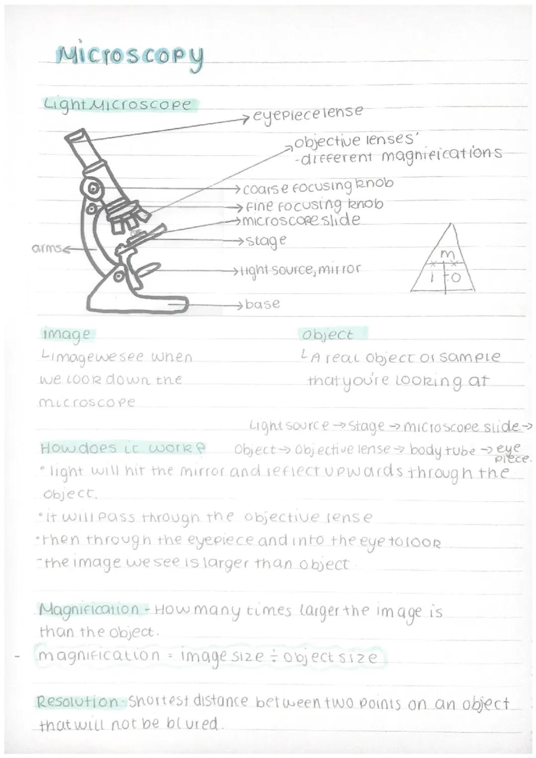
Light Microscope Basics
You'll find light microscopes in every biology lab because they're brilliant at revealing the hidden details of cells, tissues, and tiny organisms. The main parts you need to know include the eyepiece lens (where you look), objective lenses with different magnifications, and the stage where you place your sample.
The coarse and fine focusing knobs help you get crystal-clear images. Use the coarse knob first to get roughly in focus, then switch to the fine knob for that perfect, sharp view. The light source at the bottom illuminates your specimen from below.
How the magic happens: Light travels from the source, through your specimen on the microscope slide, then through the objective lens, up the body tube, and finally through the eyepiece into your eye. This clever arrangement makes tiny objects appear much larger than they actually are.
Quick Tip: Always start with the lowest magnification objective lens when examining a new specimen - it's much easier to find what you're looking for!
Understanding magnification and resolution is crucial for your exams. Magnification tells you how many times larger the image appears compared to the real object, whilst resolution determines how clearly you can distinguish between two close points.
We thought you’d never ask...
Our AI Companion is a student-focused AI tool that offers more than just answers. Built on millions of Knowunity resources, it provides relevant information, personalised study plans, quizzes, and content directly in the chat, adapting to your individual learning journey.
You can download the app from Google Play Store and Apple App Store.
That's right! Enjoy free access to study content, connect with fellow students, and get instant help – all at your fingertips.
Similar content
Most popular content: Structure of a Microscope
6Microscope Magnification Guide
Explore the essential concepts of microscope magnification, including how to calculate image size, actual size, and total magnification. This practical guide covers the preparation of microscope slides, the use of stains, and key components of light microscopes. Ideal for biology students and practical lab work.
101,04633
GCSE AQA cell biology
Grade 9 spec based revision notes for AQA higher triple bio
112722
Microscope Types Explained
Explore the key differences between light and electron microscopes, including their magnification capabilities and resolution. This summary covers essential concepts such as the structure of microscopes, the types of samples they can analyze, and how to calculate magnification. Ideal for students studying microscopy.
112913
Microscopy - introduction
light microscope parts, magnification and resolution
11510
Electron Microscopy Overview
Explore the fundamentals of electron microscopy, including Transmission Electron Microscopes (TEM) and Scanning Electron Microscopes (SEM). This summary highlights their advantages, disadvantages, and comparisons with light microscopes, focusing on resolution, magnification, and specimen preparation. Ideal for students studying microscopy techniques.
123866
Light Microscopy Techniques
Explore essential techniques for preparing and viewing specimens using light microscopy. This guide covers the components of a light microscope, specimen preparation, and practical steps for achieving clear images. Ideal for GCSE Combined Science students studying AQA and OCR Gateway. Includes practice questions to reinforce learning.
95798
Most popular content in Biology
9C
Cell Biology and Cell structure
cell structures
93,2730
T
The functions of subcellular structures - B1 Biology
Flashcards on the different functions of subcellular structures: cell membrane, nucleus, mitochondria, ribosomes, cytoplasm, permant vacuole, chloroplasts and cell wall.
102,8215
1
1.cells Gcse biology question cards
combined science higher biology
92,3604
AQA Biology: Key Concepts
Explore essential AQA Biology topics including Photosynthesis, Respiration, Homeostasis, Genetics, and Ecology. This comprehensive knowledge organizer covers key concepts such as energy transfer, hormonal control, and genetic variation, providing a solid foundation for your studies. Ideal for exam preparation and understanding biological processes.
108,851307
B
Biology Paper 1 quiz
this is a simple quiz on key knowledge needed for paper 1
102,7825
T
Types of cells
biology
101,2150
A-Level Biology Year 1 Overview
Comprehensive summary of AQA A-Level Biology Year 1, covering key topics such as cellular structure, protein synthesis, immune response, gas exchange, and more. Ideal for exam preparation and understanding biological concepts. Includes detailed insights into cellular processes, biological classification, and the circulatory system.
1214,923697
biology paper 1
all notes
102,47037
B
biology paper 1
these are some exam questions for biology paper which are more likely to come up in exams
101,8030
Most popular content
9Sociology of Education Overview
Explore comprehensive A-Level Sociology notes on the education system, covering key theories, policies, and sociological perspectives. This resource includes insights on marketisation, gender roles, cultural deprivation, and educational inequalities, providing a thorough understanding of how education shapes social stratification and individual achievement. Ideal for exam preparation and in-depth study.
12102,9323,041
Sociology of Families: Comprehensive Revision
Dive into an extensive overview of family dynamics, perspectives, and patterns in sociology. This resource covers key concepts such as family diversity, gender roles, marriage, and the impact of social policies on family structures. Perfect for A-Level Sociology students preparing for Paper 2.
1273,7402,307
Criminology: Crime & Punishment Overview
Comprehensive mindmaps covering key concepts in the Crime and Punishment topic for WJEC Criminology Unit 4. This resource includes detailed insights into the Criminal Justice System, crime prevention strategies, sentencing models, and the roles of various agencies. Ideal for A-Level revision, ensuring you grasp essential theories and legislative processes to excel in your exams.
1254,8921,060
Comprehensive Crime & Deviance Overview
Explore an extensive revision of crime and deviance topics, including theories, types of crime, and the impact of media. This resource covers key concepts such as Marxism, functionalism, gender and crime, and the influence of globalization on criminal behavior. Ideal for students seeking a thorough understanding of criminology and its various theories. Type: Full Topic Revision.
1251,6681,400
C
Cell Biology and Cell structure
cell structures
93,2730
WJEC Unit 4 Criminology
Criminology unit 4 detailed revision note
127,174125
Sociological Theories Overview
Comprehensive revision of key sociological theories including Functionalism, Marxism, Feminism, and Interpretivism. Explore concepts like value freedom, identity formation, and the critique of social control. Ideal for AQA A-Level Sociology students preparing for exams. This summary covers essential theories and their implications in sociology, providing a clear understanding of each perspective.
1231,474846
T
The functions of subcellular structures - B1 Biology
Flashcards on the different functions of subcellular structures: cell membrane, nucleus, mitochondria, ribosomes, cytoplasm, permant vacuole, chloroplasts and cell wall.
102,8215
1
1.cells Gcse biology question cards
combined science higher biology
92,3604
Students love us — and so will you.
4.6/5App Store
4.7/5Google Play
The app is very easy to use and well designed. I have found everything I was looking for so far and have been able to learn a lot from the presentations! I will definitely use the app for a class assignment! And of course it also helps a lot as an inspiration.
Stefan SiOS user
This app is really great. There are so many study notes and help [...]. My problem subject is French, for example, and the app has so many options for help. Thanks to this app, I have improved my French. I would recommend it to anyone.
Samantha KlichAndroid user
Wow, I am really amazed. I just tried the app because I've seen it advertised many times and was absolutely stunned. This app is THE HELP you want for school and above all, it offers so many things, such as workouts and fact sheets, which have been VERY helpful to me personally.
AnnaiOS user
Exploring Microscopy in Foundation Biology
Fariha Nabirah@farihanabirah
Ever wondered how scientists can see tiny cells and bacteria that are invisible to the naked eye? Light microscopes are your gateway to exploring the microscopic world, making objects appear hundreds of times larger than they actually are.
1
of 1
Sign up to see the content. It's free!
- Access to all documents
- Improve your grades
- Join milions of students
By signing up you accept Terms of Service and Privacy Policy
Light Microscope Basics
You'll find light microscopes in every biology lab because they're brilliant at revealing the hidden details of cells, tissues, and tiny organisms. The main parts you need to know include the eyepiece lens (where you look), objective lenses with different magnifications, and the stage where you place your sample.
The coarse and fine focusing knobs help you get crystal-clear images. Use the coarse knob first to get roughly in focus, then switch to the fine knob for that perfect, sharp view. The light source at the bottom illuminates your specimen from below.
How the magic happens: Light travels from the source, through your specimen on the microscope slide, then through the objective lens, up the body tube, and finally through the eyepiece into your eye. This clever arrangement makes tiny objects appear much larger than they actually are.
Quick Tip: Always start with the lowest magnification objective lens when examining a new specimen - it's much easier to find what you're looking for!
Understanding magnification and resolution is crucial for your exams. Magnification tells you how many times larger the image appears compared to the real object, whilst resolution determines how clearly you can distinguish between two close points.
We thought you’d never ask...
Our AI Companion is a student-focused AI tool that offers more than just answers. Built on millions of Knowunity resources, it provides relevant information, personalised study plans, quizzes, and content directly in the chat, adapting to your individual learning journey.
You can download the app from Google Play Store and Apple App Store.
That's right! Enjoy free access to study content, connect with fellow students, and get instant help – all at your fingertips.
Similar content
Most popular content: Structure of a Microscope
6Microscope Magnification Guide
Explore the essential concepts of microscope magnification, including how to calculate image size, actual size, and total magnification. This practical guide covers the preparation of microscope slides, the use of stains, and key components of light microscopes. Ideal for biology students and practical lab work.
101,04633
GCSE AQA cell biology
Grade 9 spec based revision notes for AQA higher triple bio
112722
Microscope Types Explained
Explore the key differences between light and electron microscopes, including their magnification capabilities and resolution. This summary covers essential concepts such as the structure of microscopes, the types of samples they can analyze, and how to calculate magnification. Ideal for students studying microscopy.
112913
Microscopy - introduction
light microscope parts, magnification and resolution
11510
Electron Microscopy Overview
Explore the fundamentals of electron microscopy, including Transmission Electron Microscopes (TEM) and Scanning Electron Microscopes (SEM). This summary highlights their advantages, disadvantages, and comparisons with light microscopes, focusing on resolution, magnification, and specimen preparation. Ideal for students studying microscopy techniques.
123866
Light Microscopy Techniques
Explore essential techniques for preparing and viewing specimens using light microscopy. This guide covers the components of a light microscope, specimen preparation, and practical steps for achieving clear images. Ideal for GCSE Combined Science students studying AQA and OCR Gateway. Includes practice questions to reinforce learning.
95798
Most popular content in Biology
9C
Cell Biology and Cell structure
cell structures
93,2730
T
The functions of subcellular structures - B1 Biology
Flashcards on the different functions of subcellular structures: cell membrane, nucleus, mitochondria, ribosomes, cytoplasm, permant vacuole, chloroplasts and cell wall.
102,8215
1
1.cells Gcse biology question cards
combined science higher biology
92,3604
AQA Biology: Key Concepts
Explore essential AQA Biology topics including Photosynthesis, Respiration, Homeostasis, Genetics, and Ecology. This comprehensive knowledge organizer covers key concepts such as energy transfer, hormonal control, and genetic variation, providing a solid foundation for your studies. Ideal for exam preparation and understanding biological processes.
108,851307
B
Biology Paper 1 quiz
this is a simple quiz on key knowledge needed for paper 1
102,7825
T
Types of cells
biology
101,2150
A-Level Biology Year 1 Overview
Comprehensive summary of AQA A-Level Biology Year 1, covering key topics such as cellular structure, protein synthesis, immune response, gas exchange, and more. Ideal for exam preparation and understanding biological concepts. Includes detailed insights into cellular processes, biological classification, and the circulatory system.
1214,923697
biology paper 1
all notes
102,47037
B
biology paper 1
these are some exam questions for biology paper which are more likely to come up in exams
101,8030
Most popular content
9Sociology of Education Overview
Explore comprehensive A-Level Sociology notes on the education system, covering key theories, policies, and sociological perspectives. This resource includes insights on marketisation, gender roles, cultural deprivation, and educational inequalities, providing a thorough understanding of how education shapes social stratification and individual achievement. Ideal for exam preparation and in-depth study.
12102,9323,041
Sociology of Families: Comprehensive Revision
Dive into an extensive overview of family dynamics, perspectives, and patterns in sociology. This resource covers key concepts such as family diversity, gender roles, marriage, and the impact of social policies on family structures. Perfect for A-Level Sociology students preparing for Paper 2.
1273,7402,307
Criminology: Crime & Punishment Overview
Comprehensive mindmaps covering key concepts in the Crime and Punishment topic for WJEC Criminology Unit 4. This resource includes detailed insights into the Criminal Justice System, crime prevention strategies, sentencing models, and the roles of various agencies. Ideal for A-Level revision, ensuring you grasp essential theories and legislative processes to excel in your exams.
1254,8921,060
Comprehensive Crime & Deviance Overview
Explore an extensive revision of crime and deviance topics, including theories, types of crime, and the impact of media. This resource covers key concepts such as Marxism, functionalism, gender and crime, and the influence of globalization on criminal behavior. Ideal for students seeking a thorough understanding of criminology and its various theories. Type: Full Topic Revision.
1251,6681,400
C
Cell Biology and Cell structure
cell structures
93,2730
WJEC Unit 4 Criminology
Criminology unit 4 detailed revision note
127,174125
Sociological Theories Overview
Comprehensive revision of key sociological theories including Functionalism, Marxism, Feminism, and Interpretivism. Explore concepts like value freedom, identity formation, and the critique of social control. Ideal for AQA A-Level Sociology students preparing for exams. This summary covers essential theories and their implications in sociology, providing a clear understanding of each perspective.
1231,474846
T
The functions of subcellular structures - B1 Biology
Flashcards on the different functions of subcellular structures: cell membrane, nucleus, mitochondria, ribosomes, cytoplasm, permant vacuole, chloroplasts and cell wall.
102,8215
1
1.cells Gcse biology question cards
combined science higher biology
92,3604
Students love us — and so will you.
4.6/5App Store
4.7/5Google Play
The app is very easy to use and well designed. I have found everything I was looking for so far and have been able to learn a lot from the presentations! I will definitely use the app for a class assignment! And of course it also helps a lot as an inspiration.
Stefan SiOS user
This app is really great. There are so many study notes and help [...]. My problem subject is French, for example, and the app has so many options for help. Thanks to this app, I have improved my French. I would recommend it to anyone.
Samantha KlichAndroid user
Wow, I am really amazed. I just tried the app because I've seen it advertised many times and was absolutely stunned. This app is THE HELP you want for school and above all, it offers so many things, such as workouts and fact sheets, which have been VERY helpful to me personally.
AnnaiOS user