Ready to tackle cell biology and microscopy? This revision guide...
Understanding Cell Structure & Key Biological Molecules for OCR A Level Biology
Ash@ash_oeqjd


1 / 10
1
of 10
Biology Revision Overview
This revision guide covers two essential biology topics that form the foundation of your A-level studies. Cell structure helps you understand how life is organised at the microscopic level, whilst biological molecules reveals the chemistry behind living processes.
The cell structure section explores everything from organelles and their functions to the key differences between prokaryotic and eukaryotic cells. You'll also master microscopy skills, including magnification calculations and comparing different types of microscopes.
The biological molecules section starts with water's vital properties before moving on to macromolecules and polymers. Carbohydrates get special attention since they're fundamental energy sources and structural components in living organisms.
Exam Tip: These topics are heavily tested, so focus on understanding functions rather than just memorising structures.
2
of 10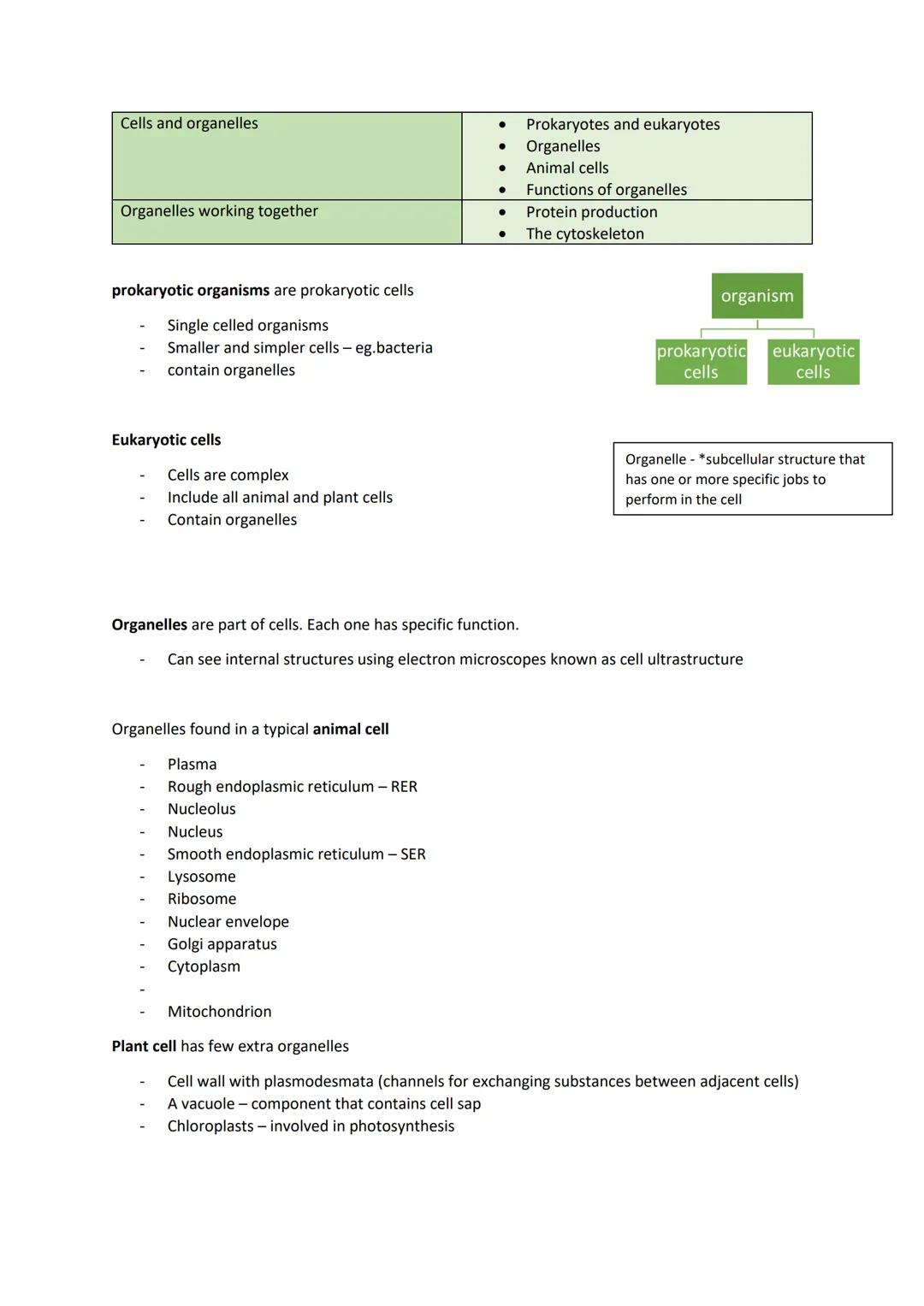
Cell Types and Basic Structures
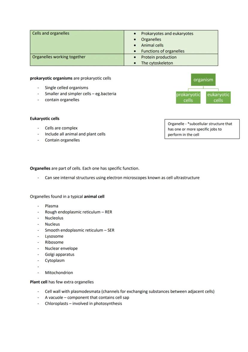
Prokaryotic cells are the simple ones - think bacteria. They're single-celled organisms that are much smaller and less complex than the cells in your body. These cells don't have a proper nucleus or membrane-bound organelles.
Eukaryotic cells are the complex ones found in animals, plants, and fungi. They're packed with organelles - specialised structures that each have specific jobs, like tiny factories within the cell. You can see their internal structures using electron microscopes, revealing what scientists call cell ultrastructure.
Animal cells contain organelles like the nucleus, mitochondria, ribosomes, and the rough endoplasmic reticulum (RER). Plant cells have all of these plus a few extras: a rigid cell wall made of cellulose, a large vacuole filled with cell sap, and chloroplasts for photosynthesis.
The plasma membrane surrounds all cells, controlling what enters and exits whilst responding to chemical signals like hormones.
Key Point: Remember that organelles are like specialised departments in a factory - each one has a specific function that keeps the cell running smoothly.
3
of 10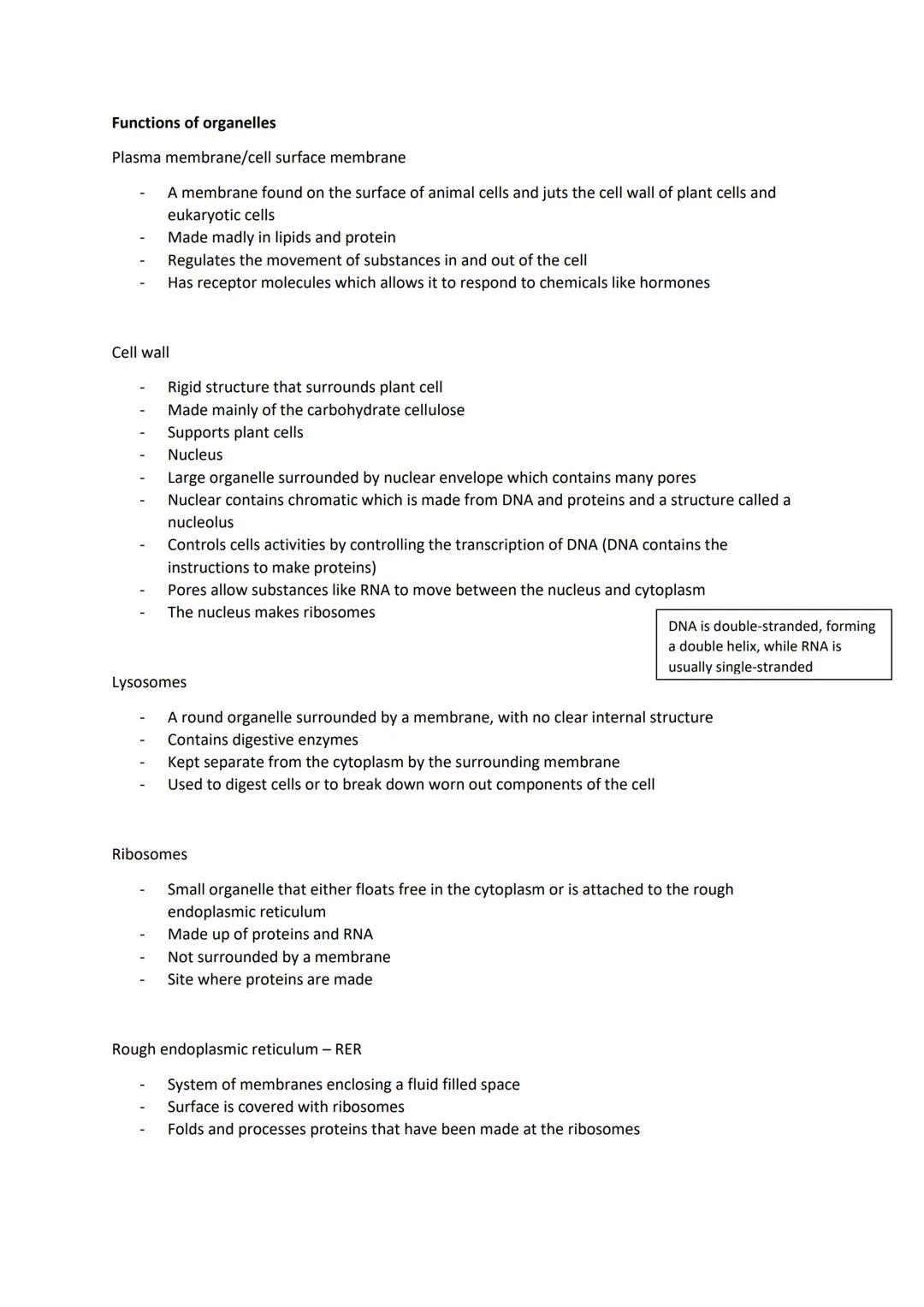
Organelle Functions
The nucleus is the cell's control centre, surrounded by a double membrane with pores. It contains chromatin (DNA plus proteins) and controls cell activities by managing protein synthesis. The nucleolus inside makes ribosomes.
Ribosomes are protein-making machines. Some float freely in the cytoplasm (making proteins that stay in the cell), whilst others attach to the rough endoplasmic reticulum (making proteins for export or membrane use).
The RER folds and processes proteins made by its attached ribosomes. The smooth endoplasmic reticulum (SER) lacks ribosomes and synthesises lipids instead. Vesicles transport materials around the cell like tiny delivery trucks.
Mitochondria are the cell's powerhouses, producing ATP through aerobic respiration. They have a double membrane with folded inner sections called cristae. Active cells need loads of mitochondria for energy. Lysosomes contain digestive enzymes to break down worn-out cell parts or digest invaders.
Memory Trick: Think of the cell as a busy factory where each organelle has a specialised job - nucleus as management, ribosomes as assembly lines, and mitochondria as the power plant.
4
of 10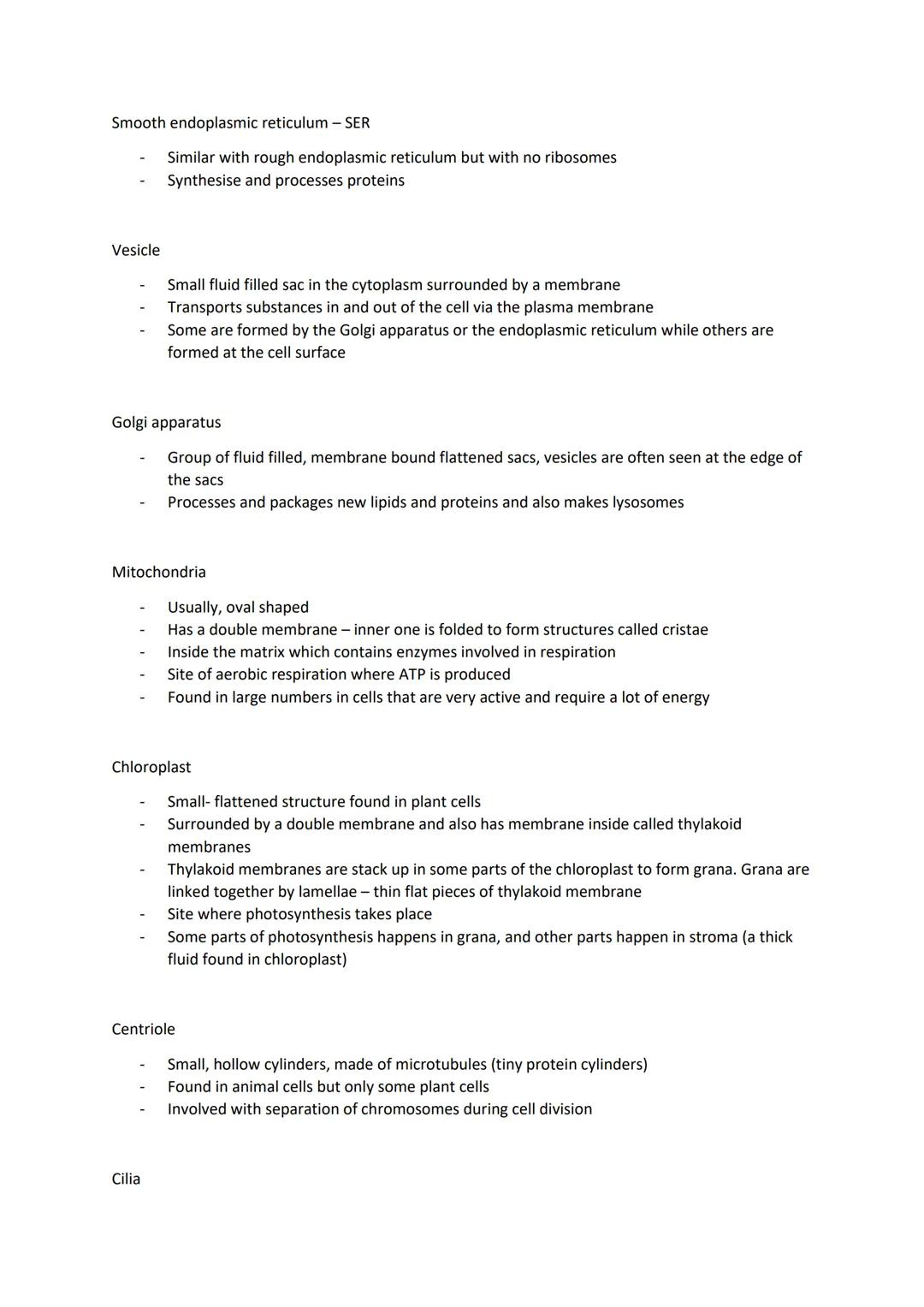
Specialised Organelles and Structures
Chloroplasts are found only in plant cells and conduct photosynthesis. They contain thylakoid membranes stacked into grana, connected by lamellae. Some photosynthesis reactions happen in the grana, others in the stroma (the thick fluid surrounding the thylakoids).
The Golgi apparatus processes and packages proteins from the ER, creating vesicles for transport. It also makes lysosomes. Think of it as the cell's post office, preparing packages for delivery.
Centrioles are hollow cylinders made of microtubules (protein tubes). They're crucial during cell division, helping separate chromosomes. Most animal cells have them, but only some plant cells do.
Cilia and flagella are hair-like projections that help cells move. Cilia are short and numerous, moving substances along cell surfaces. Flagella are longer and act like propellers - sperm cells use flagella to swim towards eggs.
Plant vs Animal: Remember that plant cells have chloroplasts, a cell wall, and a large vacuole that animal cells lack, whilst animal cells typically have centrioles that most plant cells don't.
5
of 10
Protein Production and the Cytoskeleton
Protein production involves teamwork between organelles. Ribosomes make the proteins, the RER folds and processes them, vesicles transport them to the Golgi apparatus for final processing, then more vesicles deliver the finished products where they're needed.
Proteins destined for export (like glycoproteins in mucus) follow this complete pathway from ribosomes through the RER and Golgi to the cell surface.
The cytoskeleton is a network of protein threads running through the cytoplasm. It's made of microfilaments (very thin protein strands) and microtubules (tiny protein cylinders made of tubulin).
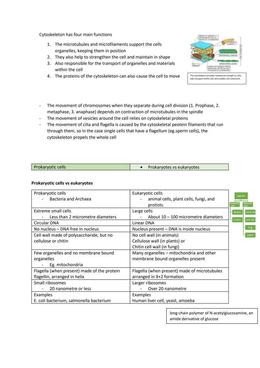
This protein network has four key functions: supporting and positioning organelles, maintaining cell shape, transporting materials within the cell, and enabling cell movement. Chromosome separation during cell division relies on microtubule contraction, and vesicle transport depends on cytoskeletal proteins.
Think of It: The cytoskeleton is like scaffolding in a building - it provides structure, support, and pathways for movement throughout the cell.
6
of 10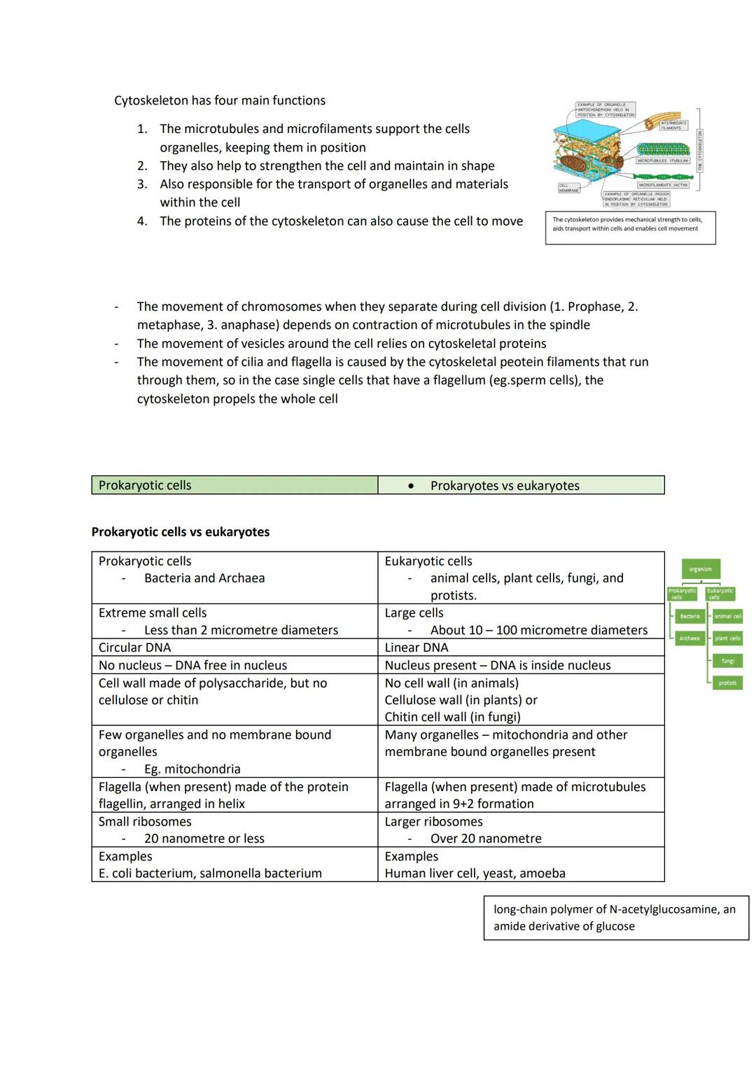
Cytoskeleton Functions and Prokaryotic Cells
The cytoskeleton drives cell movement in several ways. During cell division, microtubules contract to separate chromosomes through the phases: prophase, metaphase, and anaphase. Vesicle transport around the cell relies entirely on cytoskeletal protein tracks.
Cilia and flagella movement happens because cytoskeletal protein filaments run through them and contract. In single cells like sperm, this cytoskeletal action propels the entire cell forward.
Prokaryotic cells are much simpler than eukaryotic ones. Bacterial cells are roughly one-tenth the size of eukaryotic cells, making them difficult to study with normal microscopes.
Key bacterial structures include the bacterial chromosome (circular DNA), plasmids (small rings of DNA), cell wall, plasma membrane, and often a flagellum for movement. Unlike eukaryotic cells, bacteria lack membrane-bound organelles and a proper nucleus.
Size Matters: Prokaryotic cells are so small that you need powerful electron microscopes to see their internal details clearly.
7
of 10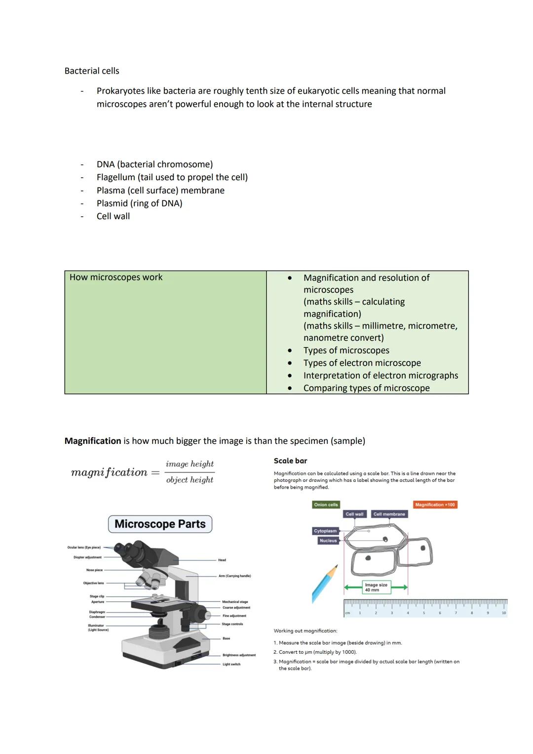
Microscopy and Magnification
Magnification tells you how much bigger an image appears compared to the actual specimen. The formula is straightforward: magnification = image height ÷ object height.
You can calculate magnification using a scale bar - that line you see on microscope photos with a measurement label. Measure the scale bar in millimetres, convert to micrometres (multiply by 1000), then divide by the actual length written on the scale bar.
Resolution is completely different from magnification - it's how much detail you can see. Think of it as the microscope's ability to distinguish between two points that are close together. If the resolution isn't good enough, just increasing magnification won't help you see more detail.
Light microscopes have a maximum resolution of about 0.2 micrometres and useful magnification up to ×1500. They're perfect for viewing whole cells and tissues. Laser scanning confocal microscopes use fluorescent dyes and can create 3D images by combining multiple scans at different depths.
Exam Focus: Practice magnification calculations - they're common exam questions and easy marks once you've got the method down.
8
of 10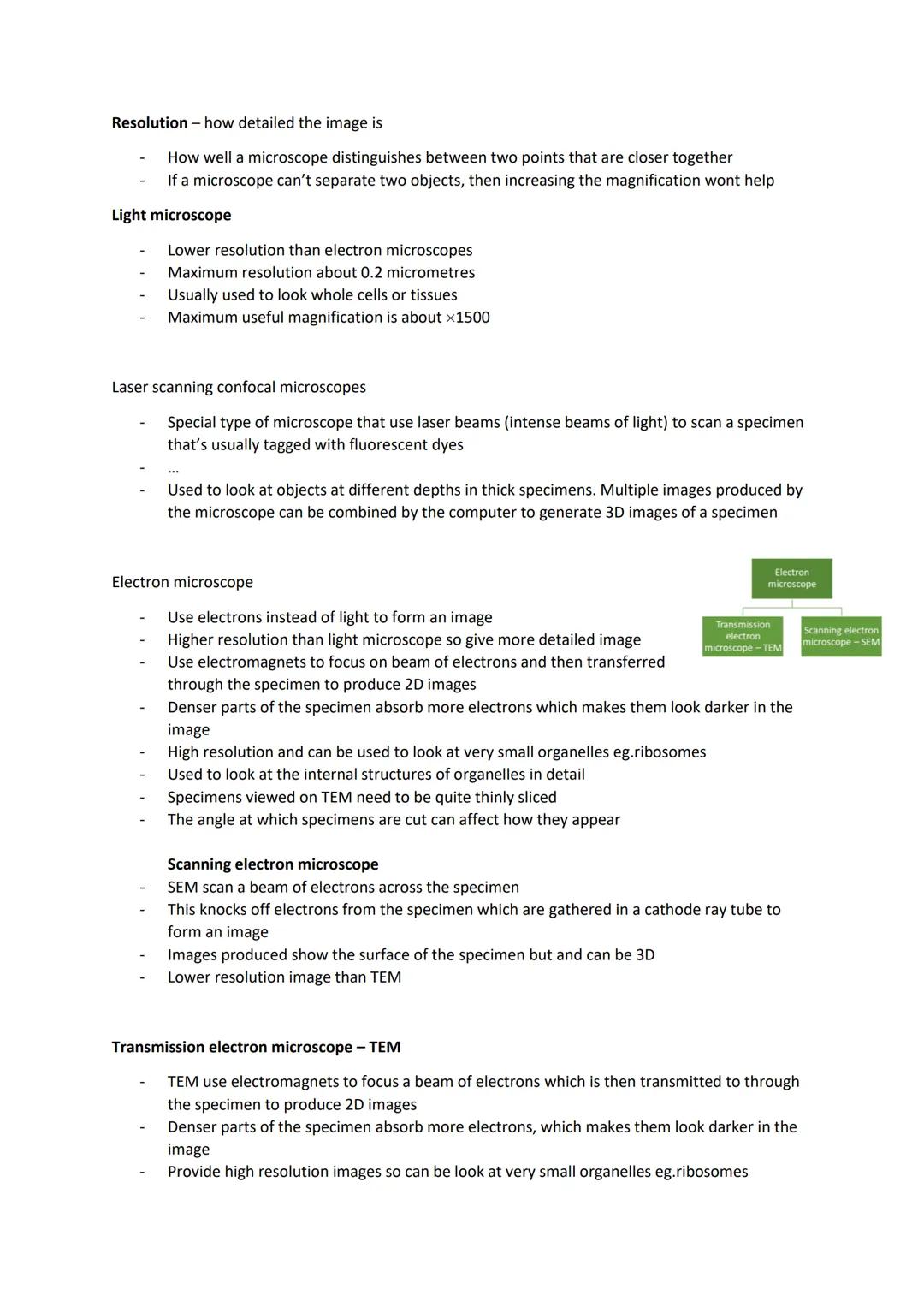
Electron Microscopes
Electron microscopes use electron beams instead of light, giving much higher resolution than light microscopes. This means incredibly detailed images of tiny structures like ribosomes and organelle interiors.
Transmission Electron Microscopes (TEMs) fire electrons through very thin specimen slices. Denser parts absorb more electrons, appearing darker in the final image. TEMs produce high-resolution 2D images perfect for studying internal organelle structures, but specimens must be sliced extremely thinly.
Scanning Electron Microscopes (SEMs) work differently - they scan electron beams across the specimen surface. Electrons get knocked off the specimen and collected to form 3D images showing surface details. However, SEM images have lower resolution than TEM images.
Both electron microscope types require specimens to be treated with heavy metals like lead. This is like staining samples for light microscopy - the metal ions scatter electrons to create contrast between different structures.
Remember: TEM = internal detail in 2D, SEM = surface detail in 3D, both give much better resolution than light microscopes.
9
of 10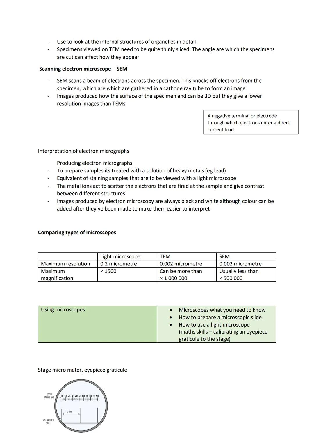
Microscope Comparison and Sample Preparation
Here's what you need to know about microscope capabilities. Light microscopes max out at 0.2 micrometre resolution and ×1500 magnification. TEMs achieve 0.002 micrometre resolution with magnification over ×1,000,000. SEMs match TEM resolution but usually stay under ×500,000 magnification.
Electron micrographs are always produced in black and white, though colours can be added later to make interpretation easier. The heavy metal treatment creates the contrast you see in these images.
For light microscopy, staining is crucial because many specimens are transparent. Methylene blue and eosin are common stains that get absorbed differently by various cell parts, creating the contrast needed to distinguish structures.
You'll use an eyepiece graticule with a stage micrometer to calibrate measurements. The stage micrometer has known measurements that help you work out the actual size of what you're viewing through the eyepiece graticule.
Practical Tip: Different stains highlight different cell components - choose your stain based on what you want to observe.
10
of 10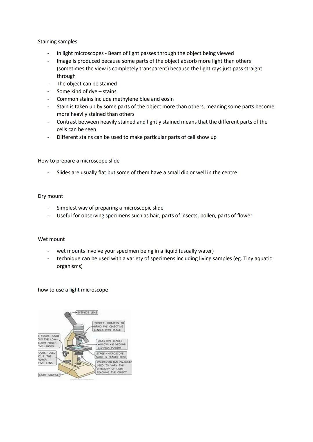
Using Light Microscopes
Staining makes transparent specimens visible by creating contrast. Some cell parts absorb more stain than others, so you get heavily stained and lightly stained areas that you can distinguish easily.
Dry mounts are the simplest slide preparation method. Just place your specimen (like hair, insect parts, or pollen) directly on the slide. Perfect for non-living samples that don't need to stay moist.
Wet mounts involve placing specimens in liquid (usually water) on the slide. This technique works brilliantly for living samples like tiny aquatic organisms that need to stay hydrated to remain active.
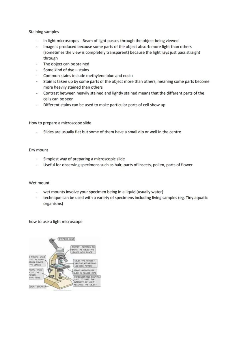
When using a light microscope, always start with the lowest power objective lens to locate your specimen, then switch to higher powers for detail. The condenser and diaphragm control light intensity reaching your specimen. Use the coarse focus for low power and fine focus for medium and high power lenses.
The turret rotates to bring different objective lenses into position. Remember that higher magnification gives a smaller field of view, so find your specimen on low power first.
Top Tip: Always start on low power and work your way up - it's much easier to find your specimen this way than starting on high magnification.
We thought you’d never ask...
Our AI Companion is a student-focused AI tool that offers more than just answers. Built on millions of Knowunity resources, it provides relevant information, personalised study plans, quizzes, and content directly in the chat, adapting to your individual learning journey.
You can download the app from Google Play Store and Apple App Store.
That's right! Enjoy free access to study content, connect with fellow students, and get instant help – all at your fingertips.
Similar content
Most popular content: Microscope
9GCSE Biology Practical Insights
Explore essential GCSE Biology practicals covering microscopy, photosynthesis, enzyme activity, food tests, and osmosis. This comprehensive guide includes key concepts, experimental procedures, and potential exam questions to enhance your understanding and preparation for Paper 1.
93,496155
Biology Required Practicals Overview
Explore the essential first four required practicals for GCSE Biology, including microscopy techniques, microbiology methods, osmosis experiments, and food testing protocols. This summary provides clear steps and safety guidelines to enhance your understanding and preparation for practical assessments.
102,48432
Microscopy Essentials
Explore the fundamentals of microscopy, including the differences between light and electron microscopes, magnification calculations, and practical usage tips. This summary covers key concepts such as microscope types, magnification formulas, and step-by-step instructions for using a light microscope, making it an essential resource for GCSE biology students.
116506
Microscope Types & Functions
Explore the various types of microscopes including Optical, Transmission Electron Microscopes (TEMs), and Scanning Electron Microscopes (SEMs). This summary covers key concepts such as magnification, resolution, and the advantages and disadvantages of each microscope type. Ideal for A-level biology students studying cell structure and microscopy techniques.
121,47138
Understanding Light Microscopes
Explore the fundamentals of light microscopes, including their components, magnification calculations, and the size range of animal and plant cells. This summary provides essential insights for biology students studying microscopy techniques and cellular structures.
11680
Types of Microscopes
Explore the differences between light and electron microscopes, including their structures, magnification capabilities, and applications in viewing cellular details. This summary provides essential insights into how these instruments enhance our understanding of microscopic life.
1144512
Microscopy Techniques Overview
Explore essential microscopy techniques for AQA A-Level Biology. This summary covers light microscopes, types of microscopes, and their applications in cell biology, tailored for Year 12 and 13 students. Enhance your understanding of microscopy with key concepts and practical insights.
111,45551
Understanding Magnification
Explore the principles of magnification in biology, focusing on how to calculate magnification using objective lens and eyepiece magnification. This summary covers key concepts such as resolution and image size, essential for Year 9-10 biology students studying microscopy and light microscopes.
91643
Microscope Practical Guide
Explore the essential techniques for observing onion and human cheek cells using a light microscope. This practical guide covers slide preparation, magnification calculations, safety measures, and a comparison between light and electron microscopes. Ideal for biology students seeking to enhance their microscopy skills.
113223
Most popular content in Biology
9C
Cell Biology and Cell structure
cell structures
93,2670
1
1.cells Gcse biology question cards
combined science higher biology
92,3584
T
The functions of subcellular structures - B1 Biology
Flashcards on the different functions of subcellular structures: cell membrane, nucleus, mitochondria, ribosomes, cytoplasm, permant vacuole, chloroplasts and cell wall.
102,8215
AQA Biology: Key Concepts
Explore essential AQA Biology topics including Photosynthesis, Respiration, Homeostasis, Genetics, and Ecology. This comprehensive knowledge organizer covers key concepts such as energy transfer, hormonal control, and genetic variation, providing a solid foundation for your studies. Ideal for exam preparation and understanding biological processes.
108,830306
A-Level Biology Year 1 Overview
Comprehensive summary of AQA A-Level Biology Year 1, covering key topics such as cellular structure, protein synthesis, immune response, gas exchange, and more. Ideal for exam preparation and understanding biological concepts. Includes detailed insights into cellular processes, biological classification, and the circulatory system.
1214,921697
B
Biology Paper 1 quiz
this is a simple quiz on key knowledge needed for paper 1
102,7795
T
Types of cells
biology
101,2120
C
Cells part 1 function of cells.
About cells and function of cells etc.
119861
B
biology paper 1
these are some exam questions for biology paper which are more likely to come up in exams
101,8010
Most popular content
9Sociology of Education Overview
Explore comprehensive A-Level Sociology notes on the education system, covering key theories, policies, and sociological perspectives. This resource includes insights on marketisation, gender roles, cultural deprivation, and educational inequalities, providing a thorough understanding of how education shapes social stratification and individual achievement. Ideal for exam preparation and in-depth study.
12102,9243,041
Sociology of Families: Comprehensive Revision
Dive into an extensive overview of family dynamics, perspectives, and patterns in sociology. This resource covers key concepts such as family diversity, gender roles, marriage, and the impact of social policies on family structures. Perfect for A-Level Sociology students preparing for Paper 2.
1273,7142,307
Criminology: Crime & Punishment Overview
Comprehensive mindmaps covering key concepts in the Crime and Punishment topic for WJEC Criminology Unit 4. This resource includes detailed insights into the Criminal Justice System, crime prevention strategies, sentencing models, and the roles of various agencies. Ideal for A-Level revision, ensuring you grasp essential theories and legislative processes to excel in your exams.
1254,8861,059
Comprehensive Crime & Deviance Overview
Explore an extensive revision of crime and deviance topics, including theories, types of crime, and the impact of media. This resource covers key concepts such as Marxism, functionalism, gender and crime, and the influence of globalization on criminal behavior. Ideal for students seeking a thorough understanding of criminology and its various theories. Type: Full Topic Revision.
1251,6671,400
C
Cell Biology and Cell structure
cell structures
93,2670
WJEC Unit 4 Criminology
Criminology unit 4 detailed revision note
127,168125
An Inspector Calls: Character Insights
Explore in-depth analysis and key quotes for characters in J.B. Priestley's 'An Inspector Calls'. This resource covers Gerald Croft, Inspector Goole, Sheila Birling, Mrs. Birling, Eric Birling, and Eva Smith, focusing on themes of class, gender roles, and social responsibility. Ideal for students aiming for Grade 8 and above.
1025,444907
Criminology Theories Overview
Explore key criminology theories and their implications on crime and deviance. This comprehensive summary covers biological, psychological, and sociological perspectives, including labelling theory, right realism, and the impact of social campaigns on policy development. Ideal for A-Level criminology students seeking to understand the complexities of criminal behaviour and the factors influencing crime prevention strategies.
129,768210
Sociological Theories Overview
Comprehensive revision of key sociological theories including Functionalism, Marxism, Feminism, and Interpretivism. Explore concepts like value freedom, identity formation, and the critique of social control. Ideal for AQA A-Level Sociology students preparing for exams. This summary covers essential theories and their implications in sociology, providing a clear understanding of each perspective.
1231,471846
Students love us — and so will you.
4.6/5App Store
4.7/5Google Play
The app is very easy to use and well designed. I have found everything I was looking for so far and have been able to learn a lot from the presentations! I will definitely use the app for a class assignment! And of course it also helps a lot as an inspiration.
Stefan SiOS user
This app is really great. There are so many study notes and help [...]. My problem subject is French, for example, and the app has so many options for help. Thanks to this app, I have improved my French. I would recommend it to anyone.
Samantha KlichAndroid user
Wow, I am really amazed. I just tried the app because I've seen it advertised many times and was absolutely stunned. This app is THE HELP you want for school and above all, it offers so many things, such as workouts and fact sheets, which have been VERY helpful to me personally.
AnnaiOS user
Understanding Cell Structure & Key Biological Molecules for OCR A Level Biology
Ash@ash_oeqjd
Ready to tackle cell biology and microscopy? This revision guide covers everything you need to know about cell structure, organelles, and the microscopes that help us see them. From the basic differences between prokaryotic and eukaryotic cells to calculating magnification...
1
of 10
Sign up to see the content. It's free!
- Access to all documents
- Improve your grades
- Join milions of students
By signing up you accept Terms of Service and Privacy Policy
Biology Revision Overview
This revision guide covers two essential biology topics that form the foundation of your A-level studies. Cell structure helps you understand how life is organised at the microscopic level, whilst biological molecules reveals the chemistry behind living processes.
The cell structure section explores everything from organelles and their functions to the key differences between prokaryotic and eukaryotic cells. You'll also master microscopy skills, including magnification calculations and comparing different types of microscopes.
The biological molecules section starts with water's vital properties before moving on to macromolecules and polymers. Carbohydrates get special attention since they're fundamental energy sources and structural components in living organisms.
Exam Tip: These topics are heavily tested, so focus on understanding functions rather than just memorising structures.
2
of 10Sign up to see the content. It's free!
- Access to all documents
- Improve your grades
- Join milions of students
By signing up you accept Terms of Service and Privacy Policy
Cell Types and Basic Structures
Prokaryotic cells are the simple ones - think bacteria. They're single-celled organisms that are much smaller and less complex than the cells in your body. These cells don't have a proper nucleus or membrane-bound organelles.
Eukaryotic cells are the complex ones found in animals, plants, and fungi. They're packed with organelles - specialised structures that each have specific jobs, like tiny factories within the cell. You can see their internal structures using electron microscopes, revealing what scientists call cell ultrastructure.
Animal cells contain organelles like the nucleus, mitochondria, ribosomes, and the rough endoplasmic reticulum (RER). Plant cells have all of these plus a few extras: a rigid cell wall made of cellulose, a large vacuole filled with cell sap, and chloroplasts for photosynthesis.
The plasma membrane surrounds all cells, controlling what enters and exits whilst responding to chemical signals like hormones.
Key Point: Remember that organelles are like specialised departments in a factory - each one has a specific function that keeps the cell running smoothly.
3
of 10Sign up to see the content. It's free!
- Access to all documents
- Improve your grades
- Join milions of students
By signing up you accept Terms of Service and Privacy Policy
Organelle Functions
The nucleus is the cell's control centre, surrounded by a double membrane with pores. It contains chromatin (DNA plus proteins) and controls cell activities by managing protein synthesis. The nucleolus inside makes ribosomes.
Ribosomes are protein-making machines. Some float freely in the cytoplasm (making proteins that stay in the cell), whilst others attach to the rough endoplasmic reticulum (making proteins for export or membrane use).
The RER folds and processes proteins made by its attached ribosomes. The smooth endoplasmic reticulum (SER) lacks ribosomes and synthesises lipids instead. Vesicles transport materials around the cell like tiny delivery trucks.
Mitochondria are the cell's powerhouses, producing ATP through aerobic respiration. They have a double membrane with folded inner sections called cristae. Active cells need loads of mitochondria for energy. Lysosomes contain digestive enzymes to break down worn-out cell parts or digest invaders.
Memory Trick: Think of the cell as a busy factory where each organelle has a specialised job - nucleus as management, ribosomes as assembly lines, and mitochondria as the power plant.
4
of 10Sign up to see the content. It's free!
- Access to all documents
- Improve your grades
- Join milions of students
By signing up you accept Terms of Service and Privacy Policy
Specialised Organelles and Structures
Chloroplasts are found only in plant cells and conduct photosynthesis. They contain thylakoid membranes stacked into grana, connected by lamellae. Some photosynthesis reactions happen in the grana, others in the stroma (the thick fluid surrounding the thylakoids).
The Golgi apparatus processes and packages proteins from the ER, creating vesicles for transport. It also makes lysosomes. Think of it as the cell's post office, preparing packages for delivery.
Centrioles are hollow cylinders made of microtubules (protein tubes). They're crucial during cell division, helping separate chromosomes. Most animal cells have them, but only some plant cells do.
Cilia and flagella are hair-like projections that help cells move. Cilia are short and numerous, moving substances along cell surfaces. Flagella are longer and act like propellers - sperm cells use flagella to swim towards eggs.
Plant vs Animal: Remember that plant cells have chloroplasts, a cell wall, and a large vacuole that animal cells lack, whilst animal cells typically have centrioles that most plant cells don't.
5
of 10Sign up to see the content. It's free!
- Access to all documents
- Improve your grades
- Join milions of students
By signing up you accept Terms of Service and Privacy Policy
Protein Production and the Cytoskeleton
Protein production involves teamwork between organelles. Ribosomes make the proteins, the RER folds and processes them, vesicles transport them to the Golgi apparatus for final processing, then more vesicles deliver the finished products where they're needed.
Proteins destined for export (like glycoproteins in mucus) follow this complete pathway from ribosomes through the RER and Golgi to the cell surface.
The cytoskeleton is a network of protein threads running through the cytoplasm. It's made of microfilaments (very thin protein strands) and microtubules (tiny protein cylinders made of tubulin).
This protein network has four key functions: supporting and positioning organelles, maintaining cell shape, transporting materials within the cell, and enabling cell movement. Chromosome separation during cell division relies on microtubule contraction, and vesicle transport depends on cytoskeletal proteins.
Think of It: The cytoskeleton is like scaffolding in a building - it provides structure, support, and pathways for movement throughout the cell.
6
of 10Sign up to see the content. It's free!
- Access to all documents
- Improve your grades
- Join milions of students
By signing up you accept Terms of Service and Privacy Policy
Cytoskeleton Functions and Prokaryotic Cells
The cytoskeleton drives cell movement in several ways. During cell division, microtubules contract to separate chromosomes through the phases: prophase, metaphase, and anaphase. Vesicle transport around the cell relies entirely on cytoskeletal protein tracks.
Cilia and flagella movement happens because cytoskeletal protein filaments run through them and contract. In single cells like sperm, this cytoskeletal action propels the entire cell forward.
Prokaryotic cells are much simpler than eukaryotic ones. Bacterial cells are roughly one-tenth the size of eukaryotic cells, making them difficult to study with normal microscopes.
Key bacterial structures include the bacterial chromosome (circular DNA), plasmids (small rings of DNA), cell wall, plasma membrane, and often a flagellum for movement. Unlike eukaryotic cells, bacteria lack membrane-bound organelles and a proper nucleus.
Size Matters: Prokaryotic cells are so small that you need powerful electron microscopes to see their internal details clearly.
7
of 10Sign up to see the content. It's free!
- Access to all documents
- Improve your grades
- Join milions of students
By signing up you accept Terms of Service and Privacy Policy
Microscopy and Magnification
Magnification tells you how much bigger an image appears compared to the actual specimen. The formula is straightforward: magnification = image height ÷ object height.
You can calculate magnification using a scale bar - that line you see on microscope photos with a measurement label. Measure the scale bar in millimetres, convert to micrometres (multiply by 1000), then divide by the actual length written on the scale bar.
Resolution is completely different from magnification - it's how much detail you can see. Think of it as the microscope's ability to distinguish between two points that are close together. If the resolution isn't good enough, just increasing magnification won't help you see more detail.
Light microscopes have a maximum resolution of about 0.2 micrometres and useful magnification up to ×1500. They're perfect for viewing whole cells and tissues. Laser scanning confocal microscopes use fluorescent dyes and can create 3D images by combining multiple scans at different depths.
Exam Focus: Practice magnification calculations - they're common exam questions and easy marks once you've got the method down.
8
of 10Sign up to see the content. It's free!
- Access to all documents
- Improve your grades
- Join milions of students
By signing up you accept Terms of Service and Privacy Policy
Electron Microscopes
Electron microscopes use electron beams instead of light, giving much higher resolution than light microscopes. This means incredibly detailed images of tiny structures like ribosomes and organelle interiors.
Transmission Electron Microscopes (TEMs) fire electrons through very thin specimen slices. Denser parts absorb more electrons, appearing darker in the final image. TEMs produce high-resolution 2D images perfect for studying internal organelle structures, but specimens must be sliced extremely thinly.
Scanning Electron Microscopes (SEMs) work differently - they scan electron beams across the specimen surface. Electrons get knocked off the specimen and collected to form 3D images showing surface details. However, SEM images have lower resolution than TEM images.
Both electron microscope types require specimens to be treated with heavy metals like lead. This is like staining samples for light microscopy - the metal ions scatter electrons to create contrast between different structures.
Remember: TEM = internal detail in 2D, SEM = surface detail in 3D, both give much better resolution than light microscopes.
9
of 10Sign up to see the content. It's free!
- Access to all documents
- Improve your grades
- Join milions of students
By signing up you accept Terms of Service and Privacy Policy
Microscope Comparison and Sample Preparation
Here's what you need to know about microscope capabilities. Light microscopes max out at 0.2 micrometre resolution and ×1500 magnification. TEMs achieve 0.002 micrometre resolution with magnification over ×1,000,000. SEMs match TEM resolution but usually stay under ×500,000 magnification.
Electron micrographs are always produced in black and white, though colours can be added later to make interpretation easier. The heavy metal treatment creates the contrast you see in these images.
For light microscopy, staining is crucial because many specimens are transparent. Methylene blue and eosin are common stains that get absorbed differently by various cell parts, creating the contrast needed to distinguish structures.
You'll use an eyepiece graticule with a stage micrometer to calibrate measurements. The stage micrometer has known measurements that help you work out the actual size of what you're viewing through the eyepiece graticule.
Practical Tip: Different stains highlight different cell components - choose your stain based on what you want to observe.
10
of 10Sign up to see the content. It's free!
- Access to all documents
- Improve your grades
- Join milions of students
By signing up you accept Terms of Service and Privacy Policy
Using Light Microscopes
Staining makes transparent specimens visible by creating contrast. Some cell parts absorb more stain than others, so you get heavily stained and lightly stained areas that you can distinguish easily.
Dry mounts are the simplest slide preparation method. Just place your specimen (like hair, insect parts, or pollen) directly on the slide. Perfect for non-living samples that don't need to stay moist.
Wet mounts involve placing specimens in liquid (usually water) on the slide. This technique works brilliantly for living samples like tiny aquatic organisms that need to stay hydrated to remain active.
When using a light microscope, always start with the lowest power objective lens to locate your specimen, then switch to higher powers for detail. The condenser and diaphragm control light intensity reaching your specimen. Use the coarse focus for low power and fine focus for medium and high power lenses.
The turret rotates to bring different objective lenses into position. Remember that higher magnification gives a smaller field of view, so find your specimen on low power first.
Top Tip: Always start on low power and work your way up - it's much easier to find your specimen this way than starting on high magnification.
We thought you’d never ask...
Our AI Companion is a student-focused AI tool that offers more than just answers. Built on millions of Knowunity resources, it provides relevant information, personalised study plans, quizzes, and content directly in the chat, adapting to your individual learning journey.
You can download the app from Google Play Store and Apple App Store.
That's right! Enjoy free access to study content, connect with fellow students, and get instant help – all at your fingertips.
Similar content
Most popular content: Microscope
9GCSE Biology Practical Insights
Explore essential GCSE Biology practicals covering microscopy, photosynthesis, enzyme activity, food tests, and osmosis. This comprehensive guide includes key concepts, experimental procedures, and potential exam questions to enhance your understanding and preparation for Paper 1.
93,496155
Biology Required Practicals Overview
Explore the essential first four required practicals for GCSE Biology, including microscopy techniques, microbiology methods, osmosis experiments, and food testing protocols. This summary provides clear steps and safety guidelines to enhance your understanding and preparation for practical assessments.
102,48432
Microscopy Essentials
Explore the fundamentals of microscopy, including the differences between light and electron microscopes, magnification calculations, and practical usage tips. This summary covers key concepts such as microscope types, magnification formulas, and step-by-step instructions for using a light microscope, making it an essential resource for GCSE biology students.
116506
Microscope Types & Functions
Explore the various types of microscopes including Optical, Transmission Electron Microscopes (TEMs), and Scanning Electron Microscopes (SEMs). This summary covers key concepts such as magnification, resolution, and the advantages and disadvantages of each microscope type. Ideal for A-level biology students studying cell structure and microscopy techniques.
121,47138
Understanding Light Microscopes
Explore the fundamentals of light microscopes, including their components, magnification calculations, and the size range of animal and plant cells. This summary provides essential insights for biology students studying microscopy techniques and cellular structures.
11680
Types of Microscopes
Explore the differences between light and electron microscopes, including their structures, magnification capabilities, and applications in viewing cellular details. This summary provides essential insights into how these instruments enhance our understanding of microscopic life.
1144512
Microscopy Techniques Overview
Explore essential microscopy techniques for AQA A-Level Biology. This summary covers light microscopes, types of microscopes, and their applications in cell biology, tailored for Year 12 and 13 students. Enhance your understanding of microscopy with key concepts and practical insights.
111,45551
Understanding Magnification
Explore the principles of magnification in biology, focusing on how to calculate magnification using objective lens and eyepiece magnification. This summary covers key concepts such as resolution and image size, essential for Year 9-10 biology students studying microscopy and light microscopes.
91643
Microscope Practical Guide
Explore the essential techniques for observing onion and human cheek cells using a light microscope. This practical guide covers slide preparation, magnification calculations, safety measures, and a comparison between light and electron microscopes. Ideal for biology students seeking to enhance their microscopy skills.
113223
Most popular content in Biology
9C
Cell Biology and Cell structure
cell structures
93,2670
1
1.cells Gcse biology question cards
combined science higher biology
92,3584
T
The functions of subcellular structures - B1 Biology
Flashcards on the different functions of subcellular structures: cell membrane, nucleus, mitochondria, ribosomes, cytoplasm, permant vacuole, chloroplasts and cell wall.
102,8215
AQA Biology: Key Concepts
Explore essential AQA Biology topics including Photosynthesis, Respiration, Homeostasis, Genetics, and Ecology. This comprehensive knowledge organizer covers key concepts such as energy transfer, hormonal control, and genetic variation, providing a solid foundation for your studies. Ideal for exam preparation and understanding biological processes.
108,830306
A-Level Biology Year 1 Overview
Comprehensive summary of AQA A-Level Biology Year 1, covering key topics such as cellular structure, protein synthesis, immune response, gas exchange, and more. Ideal for exam preparation and understanding biological concepts. Includes detailed insights into cellular processes, biological classification, and the circulatory system.
1214,921697
B
Biology Paper 1 quiz
this is a simple quiz on key knowledge needed for paper 1
102,7795
T
Types of cells
biology
101,2120
C
Cells part 1 function of cells.
About cells and function of cells etc.
119861
B
biology paper 1
these are some exam questions for biology paper which are more likely to come up in exams
101,8010
Most popular content
9Sociology of Education Overview
Explore comprehensive A-Level Sociology notes on the education system, covering key theories, policies, and sociological perspectives. This resource includes insights on marketisation, gender roles, cultural deprivation, and educational inequalities, providing a thorough understanding of how education shapes social stratification and individual achievement. Ideal for exam preparation and in-depth study.
12102,9243,041
Sociology of Families: Comprehensive Revision
Dive into an extensive overview of family dynamics, perspectives, and patterns in sociology. This resource covers key concepts such as family diversity, gender roles, marriage, and the impact of social policies on family structures. Perfect for A-Level Sociology students preparing for Paper 2.
1273,7142,307
Criminology: Crime & Punishment Overview
Comprehensive mindmaps covering key concepts in the Crime and Punishment topic for WJEC Criminology Unit 4. This resource includes detailed insights into the Criminal Justice System, crime prevention strategies, sentencing models, and the roles of various agencies. Ideal for A-Level revision, ensuring you grasp essential theories and legislative processes to excel in your exams.
1254,8861,059
Comprehensive Crime & Deviance Overview
Explore an extensive revision of crime and deviance topics, including theories, types of crime, and the impact of media. This resource covers key concepts such as Marxism, functionalism, gender and crime, and the influence of globalization on criminal behavior. Ideal for students seeking a thorough understanding of criminology and its various theories. Type: Full Topic Revision.
1251,6671,400
C
Cell Biology and Cell structure
cell structures
93,2670
WJEC Unit 4 Criminology
Criminology unit 4 detailed revision note
127,168125
An Inspector Calls: Character Insights
Explore in-depth analysis and key quotes for characters in J.B. Priestley's 'An Inspector Calls'. This resource covers Gerald Croft, Inspector Goole, Sheila Birling, Mrs. Birling, Eric Birling, and Eva Smith, focusing on themes of class, gender roles, and social responsibility. Ideal for students aiming for Grade 8 and above.
1025,444907
Criminology Theories Overview
Explore key criminology theories and their implications on crime and deviance. This comprehensive summary covers biological, psychological, and sociological perspectives, including labelling theory, right realism, and the impact of social campaigns on policy development. Ideal for A-Level criminology students seeking to understand the complexities of criminal behaviour and the factors influencing crime prevention strategies.
129,768210
Sociological Theories Overview
Comprehensive revision of key sociological theories including Functionalism, Marxism, Feminism, and Interpretivism. Explore concepts like value freedom, identity formation, and the critique of social control. Ideal for AQA A-Level Sociology students preparing for exams. This summary covers essential theories and their implications in sociology, providing a clear understanding of each perspective.
1231,471846
Students love us — and so will you.
4.6/5App Store
4.7/5Google Play
The app is very easy to use and well designed. I have found everything I was looking for so far and have been able to learn a lot from the presentations! I will definitely use the app for a class assignment! And of course it also helps a lot as an inspiration.
Stefan SiOS user
This app is really great. There are so many study notes and help [...]. My problem subject is French, for example, and the app has so many options for help. Thanks to this app, I have improved my French. I would recommend it to anyone.
Samantha KlichAndroid user
Wow, I am really amazed. I just tried the app because I've seen it advertised many times and was absolutely stunned. This app is THE HELP you want for school and above all, it offers so many things, such as workouts and fact sheets, which have been VERY helpful to me personally.
AnnaiOS user